
Date: June 21, 2022

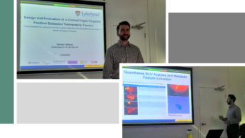
Congratulations to Justin Stiles and Brandon Baldassi as well as the other co-workers in
publishing their paper “Evaluation of a High-Sensitivity Organ-Targeted PET Camera”. Their
paper was published in the Sensors Special Issue Advanced Materials and Technologies for
Radiation Detectors.
Abstract
The aim of this study is to evaluate the performance of the Radialis organ-targeted positron
emission tomography (PET) Camera with standardized tests and through assessment of clinical-
imaging results. Sensitivity, count-rate performance, and spatial resolution were evaluated
according to the National Electrical Manufacturers Association (NEMA) NU-4 standards, with
necessary modifications to accommodate the planar detector design. The detectability of small
objects was shown with micro hotspot phantom images. The clinical performance of the camera
was also demonstrated through breast cancer images acquired with varying injected doses of 2-
[fluorine-18]-fluoro-2-deoxy-D-glucose (18F-FDG) and qualitatively compared with sample
digital full-field mammography, magnetic resonance imaging (MRI), and whole-body (WB) PET
images. Micro hotspot phantom sources were visualized down to 1.35 mm-diameter rods. Spatial
resolution was calculated to be 2.3 ± 0.1 mm for the in-plane resolution and 6.8 ± 0.1 mm for the
cross-plane resolution using maximum likelihood expectation maximization (MLEM)
reconstruction. The system peak noise equivalent count rate was 17.8 kcps at a 18F-FDG
concentration of 10.5 kBq/mL. System scatter fraction was 24%. The overall efficiency at the
peak noise equivalent count rate was 5400 cps/MBq. The maximum axial sensitivity achieved
was 3.5%, with an average system sensitivity of 2.4%. Selected results from clinical trials
demonstrate capability of imaging lesions at the chest wall and identifying false-negative X-ray
findings and false-positive MRI findings, even at up to a 10-fold dose reduction in comparison
with standard 18F-FDG doses (i.e., at 37 MBq or 1 mCi). The evaluation of the organ-targeted
Radialis PET Camera indicates that it is a promising technology for high-image-quality, low-
dose PET imaging. High-efficiency radiotracer detection also opens an opportunity to reduce
administered doses of radiopharmaceuticals and, therefore, patient exposure to radiation.
Continue reading the article: https://doi.org/10.3390/s22134678